Compassionate Care. Advanced Cardiac Treatment
CLEAR IMAGES. BETTER INSIGHT. CONFIDENT CARE.
Echocardiography
Advanced heart ultrasound imaging to assess the heart's structure, pumping function and valves, helping your cardiologist understand how your heart is working.
2D Echocardiography
Colour Doppler Assessment
Heart Function Evaluation
Detailed Cardiac Imaging
Suspected Heart Attack?
Don't Wait. Every Minute Matters.
Understanding Echocardiography
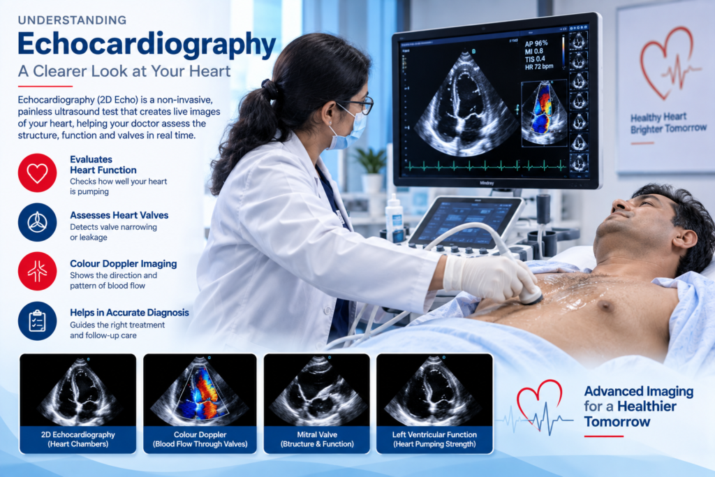
Echocardiography, commonly called an Echocardiogram or 2D Echo, is a non-invasive cardiac imaging test that uses ultrasound waves to create moving pictures of the heart. It allows the cardiologist to assess the structure and function of the heart without using X-rays.
An echocardiogram can provide information about the size and shape of the heart, how well the heart muscle pumps blood and how the heart walls move. It can also help assess the heart valves and identify problems such as narrowing or leakage of a valve.
Echocardiography may be recommended for patients with symptoms or findings that require assessment of heart function. It can be useful in evaluating conditions such as heart failure, valve disease, congenital heart conditions and after certain cardiac events, depending on the clinical situation.
Colour Doppler can be used along with echocardiography to assess the direction and pattern of blood flow through the heart and valves. The findings help the cardiologist understand the patient’s cardiac condition and decide whether further testing, treatment or follow-up is required.
Our Approach
Comprehensive Care at Every Step
Cardiac Structure Assessment
Echocardiography provides detailed information about the chambers, walls and overall structure of the heart.
Heart Pumping Function
The test helps assess how effectively the heart muscle is contracting and pumping blood.
Heart Valve Evaluation
The heart valves can be assessed for abnormalities such as narrowing or leakage and their effect on blood flow.
Colour Doppler Assessment
Doppler imaging helps evaluate blood flow through the heart and across the valves.
Monitoring Cardiac Conditions
Echocardiography may be used to assess and monitor certain heart conditions and changes in heart function over time.
Personalized Follow-up
Your cardiologist reviews the findings along with your symptoms, medical history and other investigations to determine the appropriate next step in care.
What Happens During an Echocardiogram?

Dr. Himanshu Parashar
MBBS, MD(Medicine), DNB(Cardiology), MCSI, IAMACC(America)
Before a routine transthoracic echocardiogram, usually called a 2D Echo, no major preparation is generally required. In many cases, patients can eat and drink normally unless they have been given different instructions for a specific type of echocardiogram.
During the test, you will usually lie on a couch or examination bed. Small electrodes may be placed on your chest to monitor the heart rhythm. A special gel is applied to the chest, and the sonographer moves an ultrasound probe over different areas of the chest to obtain images of the heart.
The ultrasound waves create moving images that allow the healthcare professional to assess the heart’s chambers, walls, valves and pumping function. Depending on the examination, Doppler techniques may also be used to assess blood flow through the heart and valves.
A standard echocardiogram is generally painless and does not involve radiation. The duration can vary depending on the type of examination and the information required. After the test, the images are reviewed and your cardiologist can explain the findings and whether any further evaluation or treatment is needed.
Echocardiography – Frequently Asked Questions
When is an echocardiogram recommended?
An echocardiogram may be recommended when a doctor needs to assess the heart’s structure or function. It can be useful for evaluating symptoms, heart murmurs, valve problems, heart failure and certain other cardiac conditions.
What can an echocardiogram show?
An echocardiogram can show the size and shape of the heart, heart muscle movement and pumping function, as well as the structure and function of the heart valves. It can also help identify certain abnormalities such as blood clots or structural defects.
Is a 2D Echo painful?
A routine transthoracic echocardiogram is generally painless and non-invasive. You may feel some pressure as the ultrasound probe is moved over the chest to obtain clear images.
Do I need to prepare before a 2D Echo?
For a routine transthoracic echocardiogram, special preparation is usually not required and you may generally eat and drink as usual. Preparation can be different for specialized tests such as a transesophageal echocardiogram, so follow the instructions provided by your healthcare team.
What is Colour Doppler in echocardiography?
Colour Doppler is an ultrasound technique that helps visualize blood flow within the heart and across the heart valves. It can help the cardiologist assess abnormal blood flow, including blood leaking backward through a valve or changes related to narrowed valves.
Our Location
Expert Cardiac Care Near You
- Monday to Saturday
- 10 AM to 4 PM
Supporting Better Heart Health.
Book a Cardiology Consultation